| Equine nonproliferative pododermatitis of the frog (thrush): A review S. E. O'Grady1 & T. D. Burns Reprinted with permission from Equine Veterinary Education (EVE).
SUMMARY Thrush is the term commonly used to describe nonproliferative pododermatitis of the frog epidermis caused by a bacterial infection. Thrush is commonly encountered in veterinary and farrier practices yet information on the pathogenesis, treatment and prevention of the disease in the equine veterinary literature is lacking. There is a plethora of proposed products and treatments on the market for thrush, yet most lack clinical evidence of efficacy. Clinical signs of thrush can range from black exudate in the sulci of the frog accompanied by a foul odour to bacterial invasion of the frog corium and digital cushion resulting in lameness. Thrush is primarily encountered in a conformationally compromised hoof capsule that is generally associated with an unhealthy frog. As thrush is rarely encountered in a healthy frog, it is important to focus on the farrier practices that may play a role in both the pathogenesis of thrush and its prevention. For successful treatment and prevention of thrush, good basic farrier principles must be incorporated into the treatment protocol.
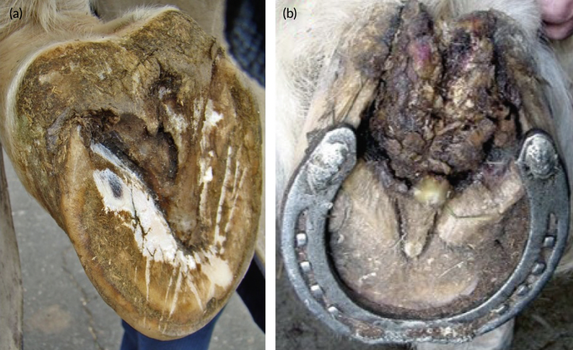
INTRODUCTION Thrush is the term commonly used to describe nonproliferative pododermatitis of the frog epidermis caused by a bacterial infection. The disease begins when keratolytic bacteria penetrate the outer horn or epidermis of the frog adjacent to the collateral sulci and/or central sulcus causing degeneration of the epidermal frog and the presence of a black necrotic exudate with a foul odour (Baxter & Stashak, 2011; Dabariener et al, 2011; O'Grady, 2020; Redding & O'Grady, 2011; Steckel, 1987). In severe cases, the bacteria can reach the underlying corium leading to significant discomfort and lameness. Blood encountered during routine hoof care when the frog is cleaned with a brush, or the end of the hoof pick is an indication the infection has extended through the epidermis of the frog into the corium and often the digital cushion. Two anaerobic bacteria species, Bacteroides sp. and Fusobacterium necrophorum, are found in equine feces/soil and are commonly associated with thrush yet published data on the true incidence of their presence in actual cases is lacking. Furthermore, both bacteria are classified as opportunistic Gram-negative pathogens with limited ability to penetrate and invade normal epidermal tissue are occasionally found which would suggest that trauma, or a compromised frog epidermis is necessary in the pathogenesis of this disease (Booth & White, 2007; Dabariener et al., 2011; Moyer, 1989). Thrush is seldom encountered in a healthy frog and it is rarely seen in the barefoot horse implying that various farriery practices may contribute to an unhealthy frog susceptible to thrush (Clayton et al., 2011; O'Grady, 2015; O'Grady & Clayton, 2023). The equine hoof capsule consists of the wall, the sole, the frog and the bulbs of the heels. The hoof capsule forms a coherent, resilient structural ‘boot’, such that distortion or disease of any given part will affect the remainder of the structures (Davies & Philip, 2007). The incidence of thrush in equine veterinary practice has been reported yet information on the pathogenesis, treatment and prevention of the disease in the equine veterinary literature is lacking. Although there is a plethora of proposed products and treatments on the market for thrush, most are anecdotal and lack clinical evidence of efficacy. Hereditary factors, the environment and unhygienic conditions, all play a role in the aetiology of this disease, but the emphasis of this review is placed on the aetiology that leads to a compromised or unhealthy frog. As thrush is rarely encountered in a healthy frog, this review will focus on the farrier practices that may play a role in both the pathogenesis of thrush and its prevention. In discussing the farriery practices implicated, the authors will use references when available as well as their clinical experience based on their combined 75 years of applying good basic farriery. Severe cases of thrush must be differentiated from canker, which is a proliferative disease where abnormal tissue (odourous filamentous fronds of hypertrophic horn—(cauliflowerlike appearance)) erodes the frog and increases in comparison to thrush, which is a degenerative disease where the horn of the frog deteriorates (O'Grady & Madison, 2004; Oosterlinck et al., 2011; Redding & O'Grady, 2011; Figure 1).
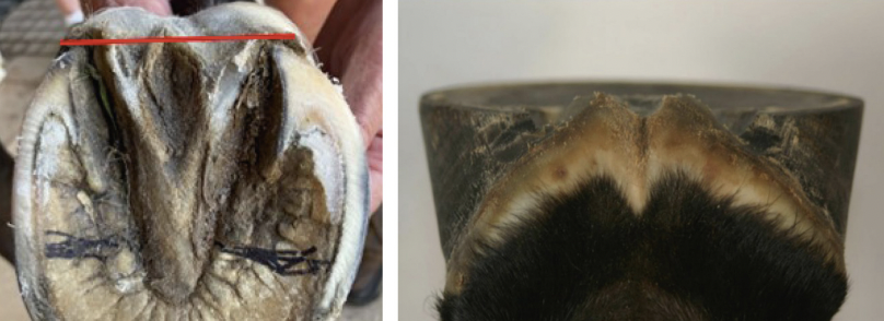
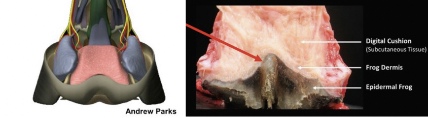
FORM AND FUNCTION OF THE FROG The frog (cuneus ungulae) is a wedge-shaped mass of keratinised stratified squamous epithelium with a softer structural texture because of an increased moisture content. Spherical masses of apocrine glands present in the corium of the frog have ducts that deliver secretions to the surface of the frog. This further contributes to the rubbery, deformable nature of the frog. The ground surface of the frog presents a pointed apex and central sulcus at the base enclosed by two crura. The central sulcus should be wide and shallow such that the index finger or ring finger fits easily into it. Paracuneal (collateral) sulci separate the crura of the frog from the bars and the sole. The palmar aspect of the frog blends into the bulbs of the heels (Figure 2). The digital cushion (pulvinus digitalis) is a highly modified subcutis consisting of a meshwork of collagenous and elastic fibres, adipose tissue, and small bundles of fibrocartilage. The digital cushion lies just proximal to the frog and frog corium, it is fixed to the adjacent structures by ligaments and thick fibre bundles. Only a few blood vessels ramify in the body of the digital cushion. It is divided into two parts: the large bilobed toric part of the digital cushion fills the bulk of the space between the heels and bulges palmarly to fill out the bulbs of the heels. The cunean part is a smaller V-shaped collagenous extension of the toric part underlying the apex of the frog. The digital cushion is composed of fat, elastic tissue, fibrous tissue and fibrocartilage cells, which are slowly dividing as only a few blood vessels ramify in the structure; therefore, it is extremely unlikely that this structure has the ability to restore or increase in size (Parks, 2011).
It is hard to separate the frog from its intimate association with the digital cushion (Figure 3). The conformation of the palmar/ plantar section which contributes to the overall conformation of the foot is determined by the size and mass of these combined structures. Furthermore, there appears to be a symbiotic relationship between these two structures with regard to function; for example, when the size of the digital cushion decreases, the mass of the frog will increase due to additional forces. This can readily be noted in the ‘bull-nosed’ low heels of the hind feet where the heels are overloaded resulting in a large bulbous frog that is weightbearing (O'Grady et al., 2018).
Function is based on the frog and digital cushion working in unison in a healthy state. If the frog fills the palmar section of the foot between the heels, contacts the ground and loads with every step during weight bearing, this distributes the load across the palmar section of the foot, and it may aid in the expansion of the hoof capsule combined with the weight of the horse (Oosterlinck et al., 2013). As part of the epidermal hoof capsule, the frog plays a major role in stabilising the hoof and provides protection for the internal structures located above. Traction is another function where the base of the frog and the descent of the apex would act as a brake on soft footing or on a slippery surface when the horse is barefoot (Davies & Philip, 2007). Obviously, some of this traction provided by the frog when barefoot would be modified by the sharp edge of the horseshoe at the toe when shod. Together with the digital cushion, the complex plays a major part in dissipating the energy of impact and concussion during the landing and stance phase of the stride (Davies & Philip, 2007). Finally, an overlooked function of the frog is to act as an expansion joint to maintain the width of the heels. If the frog loses mass and becomes recessed below the ground surface of the hoof wall at the heels or if the heels of the hoof capsule are allowed to migrate dorsally, the expansion joint is lost and the heels of the hoof capsule will start to contract and move axially (Figure 4). Furthermore, it appears that as the frog loses size and mass, the structural mass of the digital cushion also decreases; obviously, it could be deduced that a change in hoof shape results in changes in the frog. The frog and digital cushion provide a deformable interface between the rigid structures (hoof wall and ungual cartilages) of the palmar hoof capsule that permits smooth expansion and recoil during weight bearing. Although the frog and digital cushion play a predominant role in dissipating the energy of impact with the ground, it must be combined with associated soft tissue structures such as the coronary cushion, the laminar junction, the ungual cartilages and the extensive network of venous plexuses located proximal to the sole and on the abaxial surface of the ungual cartilages involved in this complex function (Davies et al., 2007).
AETIOLOGY The overall health of the frog is an important component in the aetiology of thrush. A healthy, well-formed frog is broad, firm, deformable and should fill the space between the heels of the hoof capsule. When measured at its base (the widest part), the frog width should be at least 70 per cent of its length (Turner, 1992). A healthy frog, bearing in mind that it is a deformable structure, has the ability to ‘share’ some of the load-bearing function with the other structures of the palmar/plantar hoof. In order for this to occur, the ground surface of the frog should be located on the same horizontal plane as the hoof wall at the heels. When appropriate palmar/plantar hoof conformation is present, weight bearing will stimulate the normal physiology of the frog, ensuring its continued health. A healthy frog that fills the space between the heels promotes a natural self-cleaning mechanism, such that when the foot is loaded, the frog expands, expelling any accumulated dirt or debris from the collateral frog sulci. In contrast, an unhealthy frog is markedly smaller in size and recessed below the solar surface of the hoof, thus creating a void in which debris can accumulate. Furthermore, when the frog recedes, the frog/digital cushion complex loses its ‘load sharing’ ability and weight bearing is redistributed disproportionally to the hoof wall. It appears that as the frog loses size and mass, the structural mass of the digital cushion also decreases. A compromised frog can occur for a variety of reasons including as a consequence of a recognised hoof capsule distortion such as the long toe-low heel, clubfoot or sheared heels along with inappropriate routine farriery practices. Contributing factors for thrush when the frog is not healthy include wet, unhygienic stable conditions, especially when horses continually stand in urine and manure-soiled bedding, neglecting daily routine foot care and lack of exercise (Baxter & Stashak, 2011; Dabariener et al., 2011: O'Grady, 2020; Redding & O'Grady, 2011). The fundamental problem with palmar/plantar conformation usually involves the frog not being near or on the same plane as the hoof wall at the heels and thus not contacting the ground which reduces the stimulation from the ground, causing the frog to atrophy. As the frog recedes within the hoof capsule, the frog loses its ‘self-cleaning mechanism’ which allows material (dirt/manure, etc.) to accumulate over the frog, creating excessive pressure on the frog. Over time, this constant pressure on a compromised frog leads to increased deterioration of the structures and further atrophy. Weakened by the thinning, less protective horn of the epidermis, the frog horn becomes susceptible to trauma and penetration by bacteria leading to the development of the thrush. Furthermore, the accumulation of debris over the frog creates an environment conducive to bacterial growth, especially for anaerobic organisms. The recessed frog loses its ability to act as an expansion joint and the heels begin to move axially. The diseased frog can no longer share in weight bearing, which shifts the shared function solely onto the rest of the hoof capsule. No specific organism has been identified as the sole cause of thrush. Two anaerobic bacteria species, Bacteroides sp. and Fusobacterium necrophorum are opportunistic organisms found in the soil and are commonly isolated from the bottom of the horse's foot. On the positive side, when the necrotic tissue of the frog is debrided and the structural integrity of the frog in the palmar/plantar foot is improved, the anaerobic conditions are eliminated, and bacterial growth will cease. Furthermore, if antimicrobials are required, these organisms are quite susceptible to most topical medications.
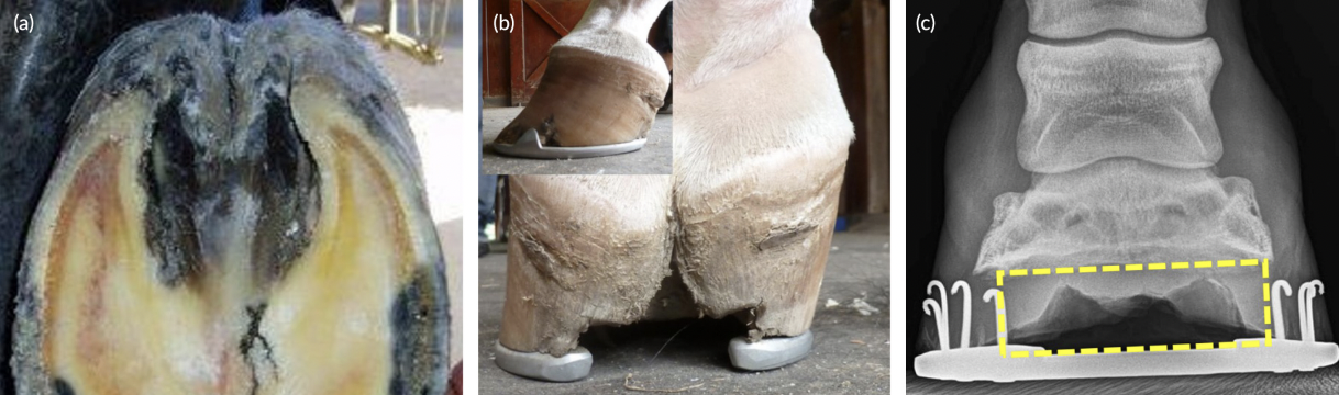
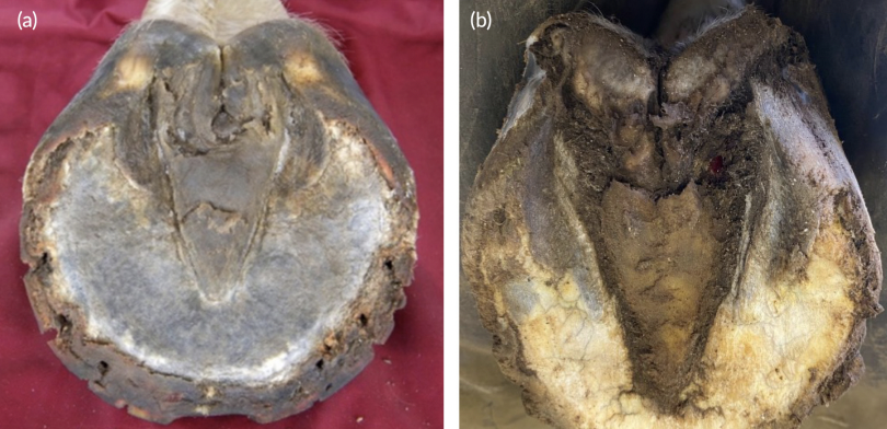
CLINICAL SIGNS AND DIAGNOSIS The diagnosis of thrush is usually straightforward and based on examination of the frog and surrounding structures. There is usually an increased amount of moisture on the bottom of the foot and a black exudate will be present in the sulci of the frog. This exudate, which varies in quantity, usually has an offensive odour. The affected sulci of the frog are often deeper than usual and may extend into the sensitive tissues of the foot causing pain. The frog will generally have decreased mass, be narrowed and recessed within the hoof capsule, lack a solid firm consistency, and have an eroded necrotic surface. There may be a fissure present in the central sulcus extending from the base of the frog to or into the hairline at the bulbs of the heels (Dabariener et al., 2011; O'Grady, 2020; Redding & O'Grady, 2011; Figure 5). The frog may also be undermined, and areas of necrotic horn are often detached from the underlying tissue. Lameness is present in severe cases that involve the dermis and swelling of the distal limb may be evident.
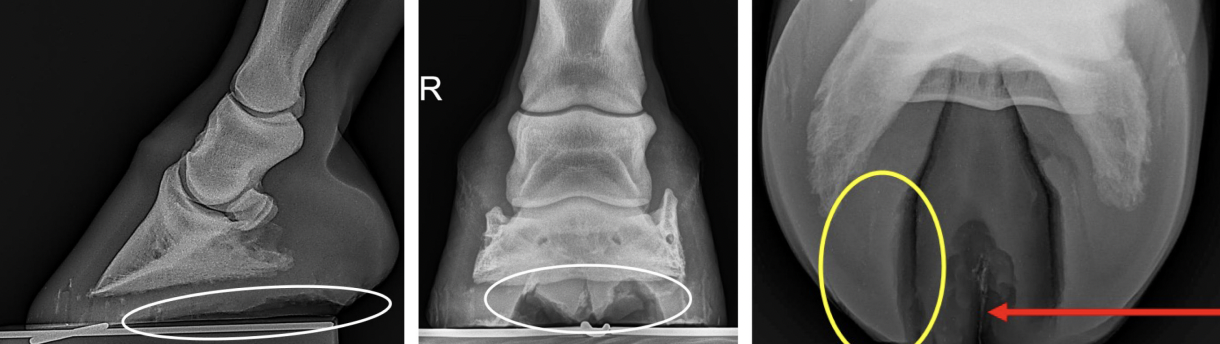
When a case of thrush is presented, the clinician should evaluate the hoof capsule to determine the cause of the compromised frog. The size and position of the frog, relative to the hoof capsule and the ground surface, should be carefully assessed. It is essential that any hoof capsule deformation such as a long toe-low heel, clubfoot or sheared heels is recognised and addressed as part of the treatment. Improving the current farriery as part of the treatment of thrush is essential. The structure of the frog and the frog fissure can readily be observed when evaluating foot radiographs. On the lateral-medial and dorso-palmar radiographs of the hoof, a lucency can be seen between the heels which indicates the frog has recessed below the ground surface of the hoof capsule. On the palmaro (45°) proximal-palmarodistal oblique or ‘skyline’ view, the outline of the frog, the frog fissure and the structures of the heels can be evaluated (Figure 6).
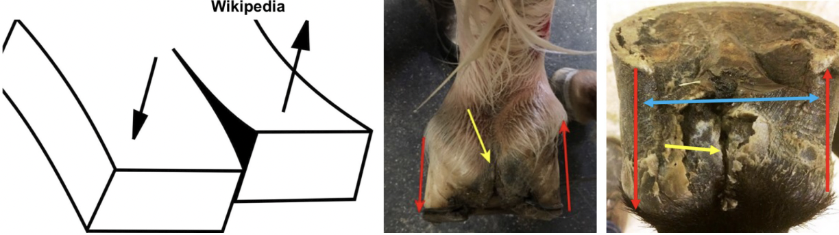
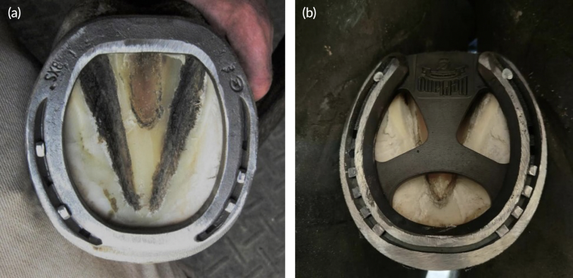
FISSURE IN CENTRAL SULCUS OF THE FROG In the unhealthy frog, a fissure will often develop in the central sulcus of the frog. This fissure or split occurring through the central sulcus has always been characterised as thrush in both the veterinary and farrier literature along with a myriad of proposed treatments. However, the authors feel the fissure is a primary structural defect that occurs in a compromised frog which creates an anaerobic environment that is ideal for the development of thrush. This fissure is often seen in horses with a hoof capsule distortion (such as low heels with structural instability or a sheared heel), a compromised frog and an abnormal strike pattern. When the frog loses structural mass and becomes recessed, the heels contract and force the central sulcus to fold inwards forming a fissure (Schumacher et al., 2012). Combined with the asymmetrical landing pattern, there is an unequal force placed on each heel causing the heels to move in opposite directions when landing. This movement in opposite directions creates a parallel shearing force on the hoof wall that leads to the formation of a full-thickness fissure between the two forces at the weakest point in the soft tissue which is the base of the frog (O'Grady, 2005, 2020; O'Grady & Castelijns, 2011; Figure 7). Significant movement in this fissure can be demonstrated with digital pressure applied by grasping a heel bulb in each hand and moving them in opposite directions. The fissure will often extend into the skin above the bulbs of the heels causing bleeding, pain and a source of lameness.
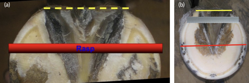
TREATMENT Treatment is divided into two subsections: farriery and medical aspects. Farriery section is necessary to understand the farrier technique used to improve the structure and integrity of the frog while the medical aspect is necessary to treat any current infection. The medical part is very basic and must be tailored to the individual case. Farriery Treatment for thrush begins with improving any hoof capsule distortion, the conformation/structures of the palmar/plantar foot, and the health of the frog. Thrush will not be resolved when the frog remains recessed below the ground surface of the foot. The amount of improvement in the frog that can be achieved is correlated with the amount of chronic damage sustained over time especially involving the frog corium. If the horse can be given time off from work, the shoes (if present) should be removed, the heels trimmed to the same horizontal plane as the frog without compromising other structures, the perimeter of the hoof wall should be strongly bevelled, and the horse left barefoot (O'Grady & Clayton, 2023). When barefoot, the load is redistributed to all the structures in the palmar foot, the integrity of structures improves rapidly and regrowth of both the frog and the heels of the hoof capsule is promoted (O'Grady, 2015; O'Grady & Clayton, 2023). Leaving the horse barefoot is very beneficial in the case with a low heel conformation where the frog has prolapsed distal to the ground surface of the foot. Here, the weight of the horse will quickly reposition the frog, thus allowing the hoof wall at the heels to be trimmed in order to create more ground surface in the palmar/plantar foot (O'Grady, 2015). Treatment of thrush while barefoot as described below is performed daily. Replacing the shoes after being barefoot or if the horse remains shod, the basic trim begins by drawing a line across the widest part of the foot and trimming the hoof wall at the heels of the hoof capsule such that the heels are on the same horizontal plane as the frog (O'Grady & Poupard, 2001) (Figure 8). This can generally be accomplished in most cases unless the frog is severely atrophied. The toe length is reduced dorsal to the line drawn across the middle of the foot, either from the bottom of the foot depending on sole thickness or from the outer dorsal hoof wall or both, resulting in approximate proportions being created on either side of the line drawn across the foot. If a significant amount of heel is removed from an upright or club foot in order to reach the desired plane, heel elevation may be necessary to accommodate for the resultant stresses placed on the deep digital flexor tendon. When a fissure is present in the central frog sulcus, it may be necessary to stabilise the heels thus decreasing the vertical movement that inhibits healing. This can readily be accomplished by applying a bar shoe or a shoe with a stabiliser plate (Figure 9). Frog pressure has and will be debated endlessly; however, it is one author's (SEO) contention, that when a flat shoe is placed on the horse's foot with good palmar soft tissue structures and the frog being on the same plane as the heels, the descent of the frog from the weight of the horse counteracted by the deformable ground surface which the horse generally encounters, places adequate but not excessive pressure on the frog. Covering the foot with a frog pad combined with silastic material is a common farriery technique in an attempt to ‘support’ the frog. However, caution should be advised as this practice may add excessive pressure and moisture on a compromised or recovering frog. Furthermore, it would impede the accessibility of the frog for daily cleaning and maintenance.
Medical Any detached areas of necrotic horn on the surface of the frog should be removed or debrided with a sharp hoof knife. The frog and adjacent area should be cleaned initially by soaking the foot in a saturated (add salt until the salt no longer dissolves) solution of Epsom salts (MgSO4). Topical antiseptics/astringents including 2% tincture of iodine, merthiolate, chlorohexidine and various copper sulfate solutions can be applied on a daily basis. Topical antibiotic ointments/solutions appear to be unnecessary. Remembering that the corium is often exposed, caustic preparations that contain phenol, formalin or formaldehyde should be discouraged. Daily aftercare Horses should have their feet cleaned and the frogs brushed vigorously with a stiff brush after which an antiseptic/astringent is reapplied on a daily basis. If there is a fissure present in the central sulcus, the area above the fissure should be flooded with the antiseptic/astringent solution to allow penetration into the fissure and then packed with cotton or a folded gauze pad. The horse should be kept in a dry, clean stall, bedded on wood shavings or sawdust and turn-out should be in a dry paddock. Exercise should be encouraged to maintain/improve the normal physiology of the foot. Repeated trimming of any detached horn on the frog may be required until the infection is controlled. Routine farriery should be scheduled every 4–5 weeks paying close attention to the trim, the foot conformation, the frog and the integrity of the soft tissue structures of the foot.
CONCLUSION There is a plethora of commercial products purported to treat/resolve thrush. However, unless the health and structural integrity of the frog is improved/restored, these products are of limited value and can perpetuate the issue. It is rare to encounter thrush in a foot with a healthy frog and support structures in the palmar/plantar foot. A review of the limited literature published on the cause and treatment of thrush does not consider the role of farriery in the aetiology of this disease process. Acquired hoof capsule distortions such as a low heel, club foot and sheared heels all affect the palmar/ plantar soft tissue structures of the foot and invariably the health of the frog. Therefore, the health of the frog cannot be improved/maintained, and consequently, the thrush cannot be resolved without combining it with the appropriate farriery. Remembering that prevention is always superior to treatment, emphasis should be placed on good basic farriery as the primary means for the prevention of equine pododermatitis.
AUTHOR CONTRIBUTIONS S. E. O'Grady: Conceptualization; investigation; writing – original draft; writing – review and editing. T. D. Burns: Writing – review and editing; methodology; investigation.
FUNDING INFORMATION There are no funders to report for this submission.
CONFLICT OF INTEREST STATEMENT No conflicts of interest have been declared.
ETHICS STATEMENT Not applicable to this review article.
ORCID S. E. O’Grady https://orcid.org/0000-0001-6243-7724
REFERENCES
| ||||||||||||||||||
| |